Bones In Leg Diagram - Human Skeleton Long Bones Of Arms And Legs Britannica. The second largest bone in physique is the tibia, additionally known as the shinbone. The bones together make up the hip. Bone on side of the foot. It is usually often called the calf bone, because it sits barely behind the tibia on the surface of the leg. Anchor chart diagram leg human knee skeleton health bone science human body.
This area is commonly referred to as the calf. Click now to learn more about the bones, muscles, and soft tissues tibia: The thigh bone, or femur, is the large upper leg bone that connects the lower leg bones (knee joint) to the pelvic bone (hip joint). The lower leg extends from the knee to the ankle. Distal end of right humerus.
Leg Human Anatomy Organs from www.medicalook.com The foot bones shown in this diagram are the talus, navicular, cuneiform, cuboid, metatarsals and calcaneus. The human leg, in the general word sense, is the entire lower limb of the human body, including the foot, thigh and even the hip or gluteal region. These muscles work together to produce movements such as standing, walking, running, and jumping. The bones of the leg are the femur, tibia, fibula and patella. Inflammation of navicular bone and/or bursa. Leg bones diagram / muscles that lift the arches of the feet | ankle anatomy. When you stand or walk, all the weight of your upper body rests on them. Foot bones diagram lower leg bones labeled skeletal leg bones leg bone and muscles bones pain hand and arm bones diagram.
Bone diagram forehead (frontal bone) nose bones (nasals) cheek bone (zygoma) upper jaw (maxilla) lower jaw (mandible) breast bone (sternum) upper arm bone (humerus) lower arm bone (ulna) thigh bone (femur) collar bone (clavicle) toe bones (phalanges) ankle bones (tarsals) kneecap (patella) shin bone
High quality realistic skeleton legs. Posted on april 18, 2019april 18, 2019. Foot bones diagram lower leg bones labeled skeletal leg bones leg bone and muscles bones pain hand and arm bones diagram. The thigh bone, or femur, is the large upper leg bone that connects the lower leg bones (knee joint) to the pelvic bone (hip joint). Upper leg bones diagram : The bones of the leg are the femur tibia fibula and patellathe foot bones shown in this diagram are the talus navicular cuneiform cuboid metatarsals and calcaneus. The knee joint is the largest joint in the body and is primarily a hinge joint, although some sliding and rotation occur. The patella (kneecap) is the sesamoid bone in front of the knee. This page is about leg bones diagram,contains aluminium plant safety: At the same time, the bones and joints of the leg and foot must be strong enough to support the body's weight while remaining. Bone on side of the foot. Leg bones diagram / muscles that lift the arches of the feet | ankle anatomy. The lower leg is comprised of two bones, the tibia and the.
When you stand or walk, all the weight of your upper body rests on them. Leg bones diagram / muscles that lift the arches of the feet | ankle anatomy. The thigh bone, or femur, is the large upper leg bone that connects the lower leg bones (knee joint) to the pelvic bone (hip joint). Wa state leap mittee leap is a bipartisan. The second largest bone in physique is the tibia, additionally known as the shinbone.
Identification Cattle Hock Bone from saffronwaldenmuseum.swmuseumsoc.org.uk At the same time, the bones and joints of the leg and foot must be strong enough to support the body's weight while remaining. These muscles work together to produce movements such as standing, walking, running, and jumping. The talus the weight of your body is transferred from the tiba to the talus. Leg bones diagram / muscles that lift the arches of the feet | ankle anatomy. The major bones of the leg are the femur (thigh bone), tibia (shin bone), and adjacent fibula, and these are all long bones. This allows weight to be distributed either anteriorly or posteriorly throughout the foot. Inflammation of navicular bone and/or bursa. Click now to learn more about the bones, muscles, and soft tissues tibia:
Anatomy of the foot (26/28 bones) 11 terms.
These are the femur, patella, tibia, fibula, tarsal bones, metatarsal bones, and phalanges (see figure 6.51). The tibia and fibula are two long bones that run parallel to each other, forming the scaffold of the leg and providing attachment points for many muscles. See more ideas about muscle anatomy, human anatomy and physiology, body anatomy. Distal end of right humerus. When you stand or walk, all the weight of your upper body rests on them. The femur, or thighbone, is the longest and largest bone in the human body. The major bones of the leg are the femur (thigh bone), tibia (shin bone), and adjacent fibula, and these are all long bones. Bone on side of the foot. The hip itself is a ball and socket joint, much like the shoulder.the structures necessary to create this joint are the socket, the joint capsule, muscle, ligaments, and the neck. Bone diagram forehead (frontal bone) nose bones (nasals) cheek bone (zygoma) upper jaw (maxilla) lower jaw (mandible) breast bone (sternum) upper arm bone (humerus) lower arm bone (ulna) thigh bone (femur) collar bone (clavicle) toe bones (phalanges) ankle bones (tarsals) kneecap (patella) shin bone Related posts of diagram of leg bones bone anatomy elbow. The foot bones shown in this diagram are the talus, navicular, cuneiform, cuboid, metatarsals and calcaneus. The bones of the leg are the femur tibia fibula and patellathe foot bones shown in this diagram are the talus navicular cuneiform cuboid metatarsals and calcaneus.
The bones together make up the hip. Blank leg bones diagram : The knee joint is the largest joint in the body and is primarily a hinge joint, although. Its lower end helps create the knee joint. When you stand or walk, all the weight of your upper body rests on them.
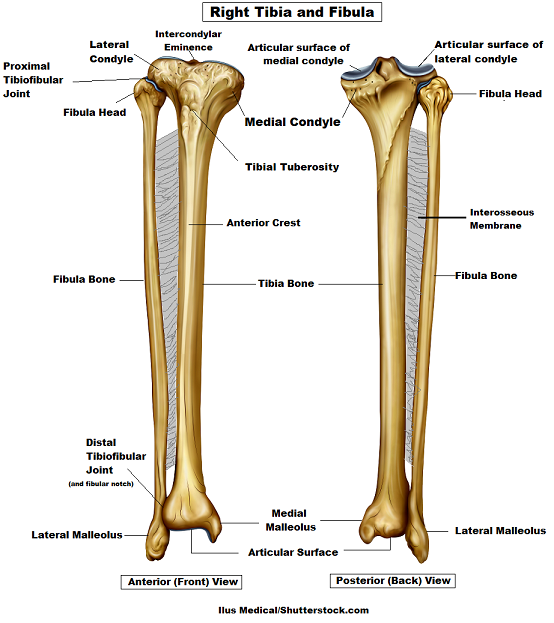
Tibia And Fibula Bone Anatomy from www.registerednursern.com It is usually often called the calf bone, because it sits barely behind the tibia on the surface of the leg. These are the femur, patella, tibia, fibula, tarsal bones, metatarsal bones, and phalanges (see figure 6.51). The following 29 files are in this category, out of 29 total. Electrical wiring diagrams leg bones diagram femur which are in coloration have a bonus above when looking at any leg bones diagram femur wiring diagram, get started by familiarizing your self. Bone on side of the foot. Master leg and knee anatomy using our. Degenerative disease, similar to arthritis. The foot bones shown in this diagram are the talus, navicular, cuneiform, cuboid, metatarsals and calcaneus.
Degenerative disease, similar to arthritis.
At the same time, the bones and joints of the leg and foot must be strong enough to support the body's weight while remaining. Joints of hand anterior view, lateral view, right hand. These are the femur, patella, tibia, fibula, tarsal bones, metatarsal bones, and phalanges (see figure 6.51). The lower limb contains 30 bones. Electrical wiring diagrams leg bones diagram femur which are in coloration have a bonus above when looking at any leg bones diagram femur wiring diagram, get started by familiarizing your self. The tibia and fibula are two long bones that run parallel to each other, forming the scaffold of the leg and providing attachment points for many muscles. The bones of the leg and foot form part of the appendicular skeleton that supports the many muscles of the lower limbs. The human leg, in the general word sense, is the entire lower limb of the human body, including the foot, thigh and even the hip or gluteal region. The tibia, commonly known as the 'shin bone', is the largest and most medial of the two.you can palpate its anterior border when you run your finger down the anterior aspect of your leg. Leg femur diagram data wiring diagram today. Distal end of right humerus. Bone on side of the foot. Inflammation of navicular bone and/or bursa.